Electrophysiological techniques are useful for studying the functional properties of cells, tissues, and organs (such as the brain and heart) with high temporal resolution. This section aims to elucidate the principles of synaptic transmission, its regulation, the functional architecture of neural circuits, and their dynamic control mechanisms, primarily through electrophysiological approaches. Moreover, we provide consultation and technical guidance on electrophysiological experiments, promote collaborative and contract research, and contribute broadly to the understanding of life phenomena. Ongoing projects are listed below.
1) Neural information processing at the tripartite synapse
Tripartite (three-part) synapses are characterized by physical connections and functional interactions between pre- and postsynaptic neurons and the surrounding glial processes. We focus on the role of neurotransmitter transporters in the integration of neuronal information at the tripartite synapse. We also analyze genetically engineered animals to understand the pathophysiology of neurological disorders, including rapid-onset dystonia with parkinsonism (RDP), alternating hemiplegia of childhood (AHC), and bipolar disorder. In addition to classical techniques such as electrophysiology, immunohistochemistry, and pharmacology, our laboratory has recently introduced photo-releasable caged compounds.
2) Regulation of neural network activity for motor learning
Neurons form complex networks between them and send information to multiple brain areas. We are investigating how neural network activity related motor learning is regulated in the cortex and the basal ganglia system (Fig. 1). We approach these questions using electrophysiology, computer simulation, and behavior analysis. We also analyze how neurotransmitters including dopamine regulate intrinsic membrane properties of cells and reward related behaviors as a research collaboration.
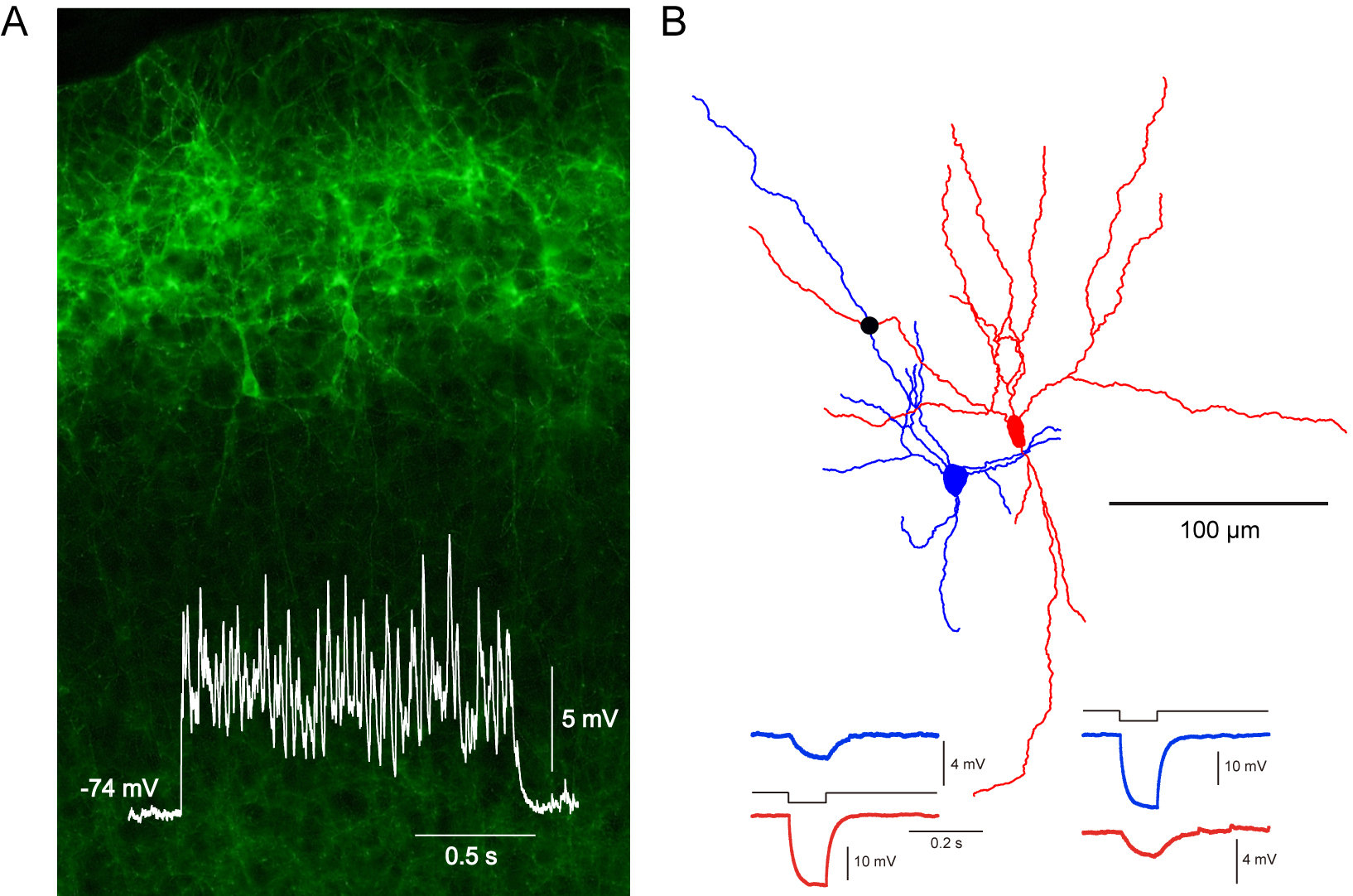
Fig.
(A) Network activity evoked by optogenetic stimulation. ChR2-Venus was selectively expressed in cortical L2/3 pyramidal cells. During light stimulation, membrane potential oscillation was induced in L5 pyramidal cell. (B) Reconstruction of cortical FS interneurons. Electrically connected FS cells, confirmed by negative current injection to one of two cells, were simultaneously recorded. ● indicates electrical connection site.


