Member
Analysis of physiological changes in multicellular circuit dynamics responsible for higher brain
1. Aim of Research
The Division Multicellular Circuit Dynamics aims to elucidate the circuitry mechanism of neuron and glia cells in central nervous system. For this purpose, 1. We focus on the glial physiological functions that affect on the neuronal circuits and ultimately on the behavior output. 2. We focus on the functional connectivity of the local multicellular circuits and quantify the connectivity by our developed holographic microscope to modulate the circuits. Please see below for the detail.
(1) Project to reveal physiological functions of glial cells
Microglia : We previously showed that microglia directly contact on synapse to monitor their functions using two photon microscopes (Wake et al., 2009). Our recent research showed that microglia contact on synapse via P2Y12 signaling to modulate their function and thus to regulate the synchronization of neuronal circuits (Akiyoshi et al., 2018, Badimon et al., 2020). In addition, we revealed that microglia migrate on blood vessels with the induction of systemic inflammation and expressed Cldn5 to form a tight junction with endothelial cells to protect their permeability. However, with the progression of inflammation, microglia start to express CD68 to phagocyte astrocyte endfeet and thus increase the BBB permeability (Haruwaka et al., 2019). In addition, somatosensory sensation is enhanced after visual deprivation (cross-modal plasticity), and we found that the neural circuits connecting the somatosensory cortex to the higher visual cortex are important for cross-modal plasticity. We also found that microglia play an important role in the circuit rewiring (Hashimoto et al., 2023). We also revealed the plasticity of microglia in early brain metastasis and highlighted their potential as early therapeutic targets (Tsuji et al., 2026).
(b) Oligodendrocyte : Activity-dependent myelination contributes to synchrony of neuronal activity and motor learning using two-photon microscopy and electrophysiological techniques (Sugio et al., submitted). In addition, the lipid synthesis is essential for activity dependent myelination and their contribution on motor learning (Kato et al., 2023).
(2) Development of holographic microscope
To manipulate neuronal and glial circuits with higher temporal and spatial resolution, we developed holographic microscope. Using this system, we measured the local circuit connectivity by stimulating single cell with simultaneous imaging of neuronal populational activity and studied the connectivity change in pain model (Okada et al., 2021). Combined with data on the plasticity of different senses, we are trying to introduce artificial sensation (Tanisumi et al., in preparation).
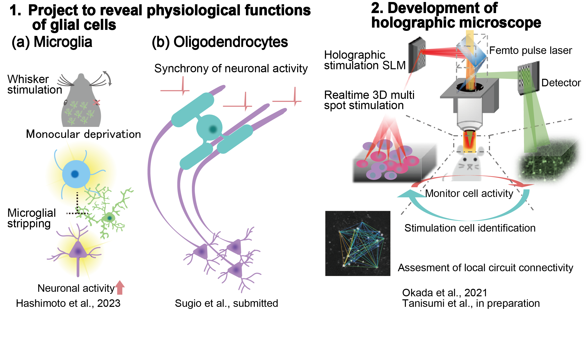
(a) Monocular deprivation induces microglial stripping of inhibitory synapses. This promotes neuronal activation of the secondary visual cortex (V2L) during whisker stimulation.
(b) Oligodendrocyte induces synchrony of neuronal activity.
Figure 2
Holographic stimulation assess the functional connectivity of local neural circuits.
Selected publications
*Akiyoshi et al., eNeuro (2018)
*Badimon et al., Nature (2020)
*Kato et al., Glia (2023)
*Haruwaka et al., Nat Commun. (2019)
*Hashimoto et al., Cell Rep. (2023)
*Okada et al., Sci Adv. (2021)
*Wake et al., J Neurosci. (2009)
*Tsuji et al., Cancer Res (2026)


