The ability to estimate the passage of time is essential for our daily life. While it has been documented that spatial features, such as orientations, are represented by the feature selective neurons that fire selectively to the specific preferred orientations, it remains largely unknown how our brain represents time intervals. The new study by Hayashi et al. used a functional magnetic resonance imaging (fMRI) adaptation paradigm to determine whether our brain has neural populations tuned to specific preferred durations. A series of experiments showed that the right supramarginal gyrus (SMG) exhibited a reduction in the fMRI signal due to adaptation when a visual stimulus of the same duration was repeatedly presented. Moreover, the fMRI adaptation was observed irrespective of the subject’s attention to time. Repetition of a nontemporal aspect of the stimulus (i.e., shape) did not produce neural adaptation in the right SMG. These results indicate that the human right SMG contains neural populations tuned to specific preferred durations.
This study was partly supported by the Cooperative Study Program of National Institute for Physiological Sciences.
Based on the findings that the right SMG showed fMRI adaptation by repetition of an identical duration, this study provides neural evidence for duration-tuned representations in the human right SMG.
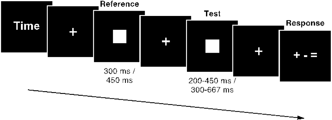 Participants performed a duration discrimination task during the fMRI scan. In this experiment (experiment 4), 300-ms reference stimulus followed by 200-, 300-, or 450-ms test stimulus, or 450-ms reference followed by 300-, 450-, or 667-ms test stimulus. Participants were asked to respond with whether the duration of the test stimulus was shorter, longer, or the same compared with the reference stimulus.
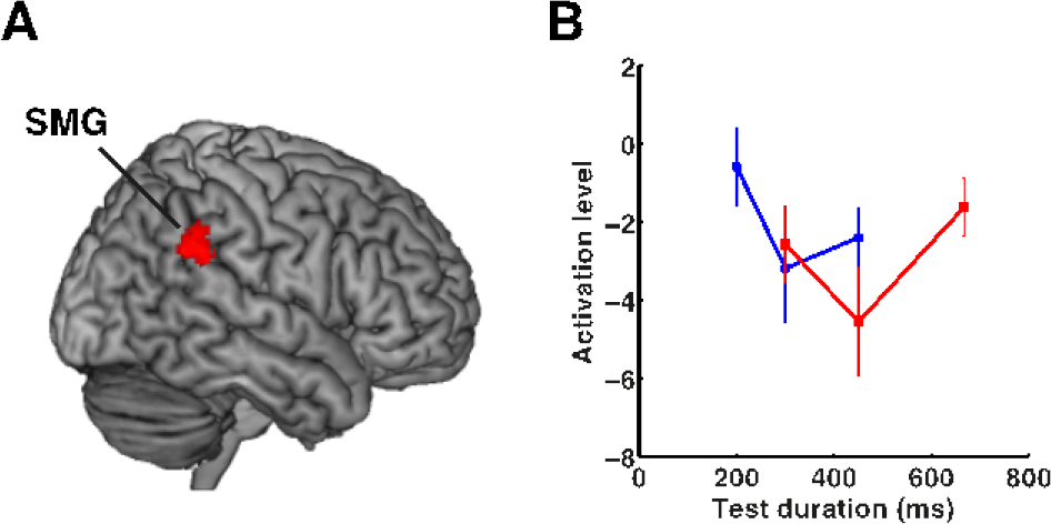
(A) The fMRI result of experiment 4. The cluster showing a duration adaptation effect was found in the right SMG. (B) Plots of the activation levels under each set of stimulus conditions in the 300-ms (blue) and the 450-ms (red) reference blocks in the right SMG. This plot clearly showed that fMRI responses were reduced when the same durations were repeated.
The present study provides new insights into the neural mechanisms of temporal estimation, learning, and prediction of future events. Deficits in temporal processing have been found in a number of neurological and psychiatric conditions, such as Parkinson’s disease, schizophrenia, attention-deficit hyperactivity disorder, and autism. Impulsive decision-making has also been linked with deficits in temporal processing. The new findings may help a better understanding of these pathological conditions and the neural mechanisms of decision-making.
Time adaptation shows duration selectivity in the human parietal cortex.
Masamichi J. Hayashi, Thomas Ditye, Tokiko Harada, Maho Hashiguchi, Norihiro Sadato, Synnöve Carlson, Vincent Walsh & Ryota Kanai.
PLOS Biology. 13(9): e1002262. September 17,2015
Dynamic Brain Network Group
Graduate School of Frontier Biosciences
Osaka University
JSPS Research Fellow
Masamichi Hayashi
Tel. +81 6 6879 4431 FAX. +81 6 6879 4437
Email: mjhgml@gmail.com


