The goal of Division of Cerebral Integration is to understand the physiology of human voluntary movement and other mental processing including language and social interaction using noninvasive functional neuroimaging technique, mainly fMRI. In particular, neural mechanisms of the social interaction is the main focus of our research activities. Hyper-scanning fMRI (3T) has been installed to evaluate two individuals as one neural network during social interaction, combined with 7T MRI data to evaluate the detailed structures of the neural network that represent the social cognition. Multimodal approach including EEG , MEG, TMS, and NIR is considered when appropriate.
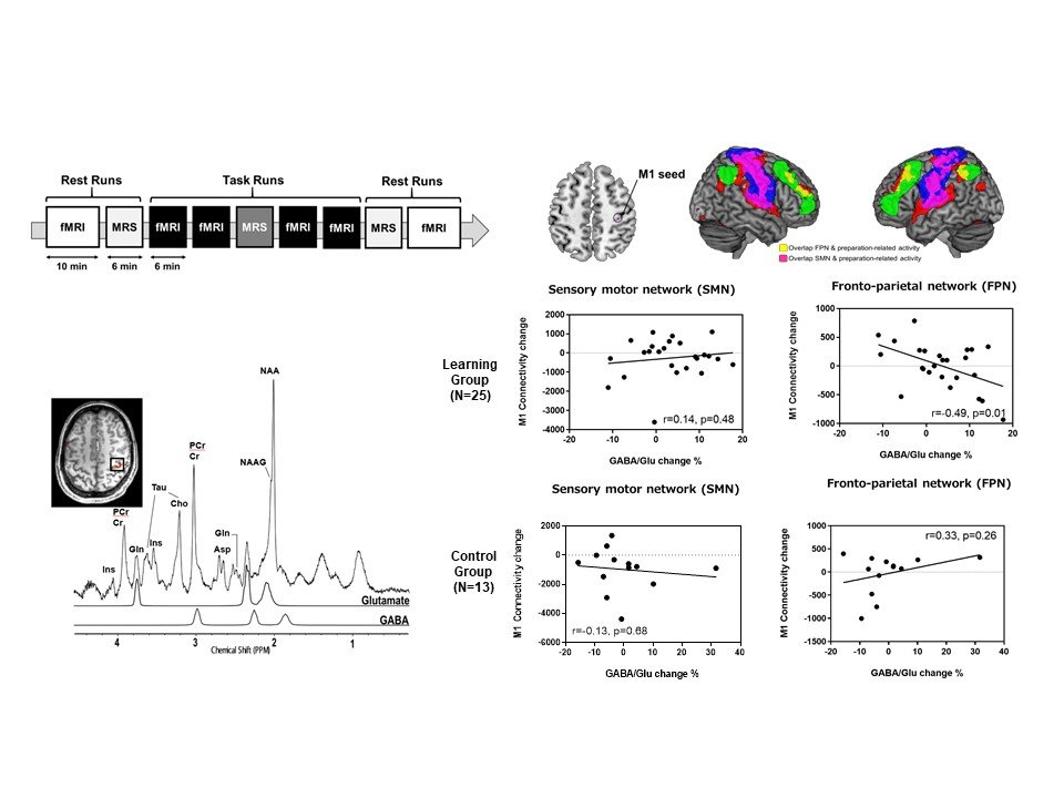
Figure 1. Neural substrates of motor engram Using 7T fMRI, we visualized the primary motor cortex (M1) encoding motor memory traces (engrams) as increased motor preparatory activity in M1 associated with learning. In addition, interactions with other brain regions during motor learning were assessed using 7TMRS in combination with fMRI during task and rest. We found cognitive control-based motor learning was associated with local changes in the GABA/glutamate ratio in M1 reflecting remote connections with the frontal-parietal executive network (FPN), which connections in turn represented motor learning memory formation at the network level.
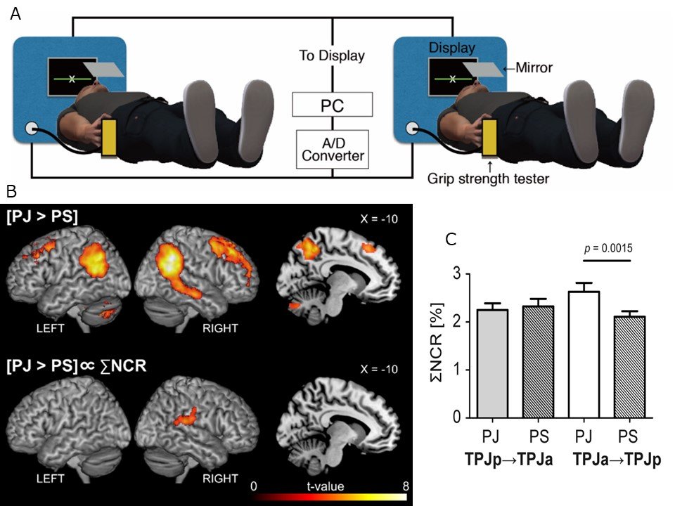
Figure 2. To evaluate the neural substrates of cooperation, we conducted a hyperscanning functional MRI study with a joint force-production task. We found that the cooperation, the degree of adjustment of individual motor output depending on that of the partner, is mediated by the interconnected subdivisions of the right TPJ.


