Shimomura T, Hirazawa K, Kubo Y (2023) Conformational rearrangements in the 2nd voltage sensor domain switch PIP2- and voltage-gating modes in two-pore channels. Proc Natl Acad Sci USA (120(6):e2209569120. doi: 10.1073/pnas.2209569120.)
Chen IS, Eldstrom J, Fedida D, Kubo Y (2022) A novel ion conducting route besides the central pore in an inherited mutant of G-protein-gated inwardly rectifying K+ channel. J Physiol 600: 603-622. doi: 10.1113/JP282430.
Andriani RT, Kubo Y (2021) Voltage-clamp fluorometry analysis of structural rearrangements of ATP-gated channel P2X2 upon hyperpolarization. Elife 10:e65822. doi: 10.7554/eLife.65822.
Division of Cardiocirculatory Signaling (Exploratory Research Center on Life and Living Systems)

Prof. NISHIDA, Motohiro

Assoc.Prof. NISHIMURA, Akiyuki
Elucidation of the molecular mechanism underlying maintenance and transfiguration of cardiovascular homeostasis
We are studying the mechanism underlying adaptation and maladaptation of the cardiovascular system against hemodynamic load through focusing on TRP Ca
2+-permeable channels as a regulator of excitation-transcription coupling and GTP-binding proteins as senescence-inducible redox sensors, using in vivo and ex vivo cardiovascular analyzing systems and chemical principles-based biological techniques.
Article Information
Nishimura A, Ogata S, Tang X, Hengphasatporn K, Umezawa K, Sanbo M, Hirabayashi M, Kato Y, Ibuki Y, Kumagai Y, Kobayashi K, Kanda Y, Urano Y, Shigeta Y, Akaike T, Nishida M (2025) Polysulfur-based bulking of dynamin-related protein 1 prevents ischemic sulfide catabolism and heart failure in mice. Nat. Commun., 16, 276
Nishimura A, Tanaka T, Shimoda K, Ida T, Sasaki S, Umezawa K, Imamura H, Urano Y, Ichinose F, Kaneko T, Akaike T, Nishida M (2025) Non-thermal atmospheric pressure plasma-irradiated cysteine protects cardiac ischemia/reperfusion injury by preserving supersulfides. Redox Biol., 79, 103445
Kato Y, Ariyoshi K, Nohara Y, Matsunaga N, Shimauchi T, Shindo N, Nishimura A, Mi X, Kim SG, Ide T, Kawanishi E, Ojida A, Nakashima N, Mori Y, Nishida M (2024) Inhibition of dynamin-related protein 1-filamin interaction improves systemic glucose metabolism. Br. J. Pharmacol., 181, 4328-4347
Oda S, Nishiyama K, Furumoto Y, Yamaguchi Y, Nishimura A, Tang XK, Kato Y, Numaga-Tomita T, Kaneko T, Mangmool S, Kuroda T, Okubo R, Sanbo M, Hirabayashi M, Sato Y, Nakagawa Y, Kuwahara K, Nagata R, Iribe G, Mori Y, Nishida M (2022) Myocardial TRPC6-mediated Zn2+ influx induces beneficial positive inotropy through β-adrenoceptors. Nat. Commun., 13, 6374
Contact: NISHIDA, Motohiro TEL:+81-564-59-5560 / E-mail:nishida(at)nips.ac.jp
NISHIMURA, Akiyuki TEL:+81-564-59-5563/ E-mail: aki(at)nips.ac.jp
Lab Website
Division of Molecular Neuroimmunology

Prof. MURAKAMI, Masaaki*
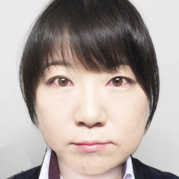
Assoc.Prof. HASEBE, Rie
Molecular mechanism of the development of autoimmune diseases regulated by gateway reflex
Prof. MURAKAMI and Assoc. Prof. HASEBE found a novel concept for neuro-immune interaction, “The Gateway Reflex”, by studying the mechanism of how IL-6 and autoreactive CD4+ T cells induce the tissue-specific autoreactive immune diseases (Cell 148, 447-, 2012, JEM 219, e20212019, 2022 etc). Six Gateway Reflexes have been found so far. In the Gateway Reflex, environmental stimulations such as gravity, pain, stress, light, and microinflammation activate the specific neural pathways, which induces accumulation of autoreactive CD4+T cells to induce inflammation around the specific blood vessels of the tissue possessing blood barrier (e.g. the central nervous system or retina). The mechanism of how the Gateway Reflex increases vascular permeability has been addressed by the concept of “IL-6 amplifier” (Immunity, 50, 812, 2019, Immunity, 29, 628-, 2008). The research goals of Division of Molecular Neuroimmunology are, 1. to discover novel Gateway Reflexes, 2. to elucidate molecular mechanism including the details of neural pathway related to the Gateway Reflexes, 3. to elucidate molecular mechanism of inflammation induced by IL-6 amplifier.
Article Information
Hasebe R, et al., (2022) ATP spreads inflammation to other limbs through crosstalk between sensory neurons and interneurons. J Exp Med 219: e20212019.
Stofkova A, et al., (2022) Depletion of Retinal Dopaminergic Activity in a Mouse Model of Rod Dysfunction Exacerbates Experimental Autoimmune Uveoretinitis: A Role for the Gateway Reflex. Int J Mol Sci 23: 453
Murakami M et al., (2019) Pleiotropy and Specificity: Insights from the interleukin 6 family of cytokines. Immunity 50: 812-831.
Stofkova A et al., (2019). Photopic light-mediated down-regulation of local α1A-adorenagic signaling protects blood-retina barrier in experimental autoimmune uveoretinitis. Sci Rep 9: 2353.
Arima Y, et al., (2017) Brain micro-inflammation at specific vessels dysregulates organ-homeostasis via the activation of a neu neural circuit. eLife 6: e25517.
Arima Y, et al., (2015) A pain-mediated neural signal induces relapse in murine autoimmune encephalomyelitis, a multiple sclerosis model. eLife 4: e08733.
Arima Y, et al., (2012) Reginal neural activation defines a gateway for autoreactive T cells to cross the blood-brain barrier. Cell 148: 447-457.
Contact: HASEBE, Rie TEL:+81-564-55-7729 / E-mail : hasebe(at)nips.ac.jp
(*Prof. MURAKAMI, Masaaki cannot accept students as a supervisor. Assoc.Prof. HASEBE, Rie accepts them instead and discusses the research with Prof. MURAKAMI, Masaaki as well.)
Lab Website
Division of Mammalian Embryogenesis
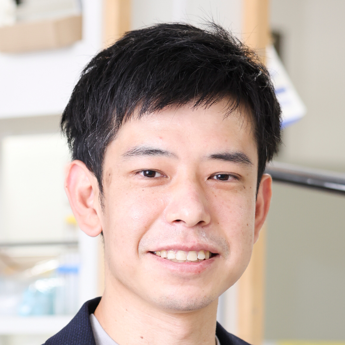
Prof. KOBAYASHI, Toshihiro
Understanding and applying developmental competency of mammalian pluripotent stem cells
We aim to understand the mechanisms underlying the developmental competency of pluripotent stem cells (PSCs) derived from early mammalian embryos and to apply their principle for in vitro gametogenesis and in vivo organ regeneration. We focus on PSCs and early embryos from various mammals, not only mice and humans, enabling us to study conserved mechanisms across species and develop novel reproductive technologies based on species-specific features.
Article Information
Iwatsuki K, Oikawa M, Kobayashi H, Penfold CA, Sanbo M, Yamamoto T, Hochi S, Kurimoto K, Hirabayashi M, Kobayashi T *. (2023) Rat post-implantation epiblast-derived pluripotent stem cells produce functional germ cells. Cell Rep Methods. Jul 27;3(8):100542.
Oikawa M*, Kobayashi H, Sanbo M, Mizuno N, Iwatsuki K, Takashima T, Yamauchi K, Yoshida F, Yamamoto T, Shinohara T, Nakauchi H, Kurimoto K, Hirabayashi M, Kobayashi T* . (2022) Functional primordial germ cell-like cells from pluripotent stem cells in rats. Science. Apr 8;376(6589):176-179.
Kobayashi T*, Castillo-Venzor A*, Penfold CA, Morgan M, Mizuno N, Tang WWC, Osada Y, Hirao M, Yoshida F, Sato H, Nakauchi H, Hirabayashi M, Surani MA *. (2021) Tracing the emergence of primordial germ cells from bilaminar disc rabbit embryos and pluripotent stem cells. Cell Rep. 2021 Oct 12;37(2):109812
Contact: KOBAYASHI, Toshihiro TEL: +81-564-59-5265 / E-mail: tkoba(at)nips.ac.jp
Lab Website
Division of Visual Information Processing
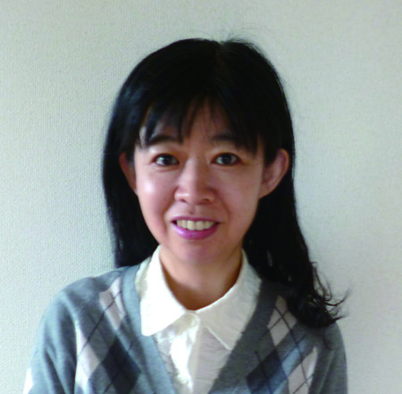
Prof. YOSHIMURA, Yumiko
Circuit mechanisms underlying information processing and functional development in visual cortex
We are studying the properties of neuronal circuits underlying visual information processing in visual cortex, and the development of the circuits based on experience-dependent and synaptic target recognition mechanisms. To this end, we are conducting electrophysiological analyses combined with optogenetics and laser photolysis of caged glutamate in rodent slice preparations and morphological analyses using transsynaptic viral tracers. In order to relate the properties of neuronal circuits to visual functions, we are also investigating the visual responses of cortical neurons in rodents with electrophysiological techniques and 2-photon Ca
2+ imaging.
Article Information
Yoneda T, Hayashi K, Yoshimura Y. Experience-dependent functional plasticity and visual response selectivity of surviving subplate neurons in mouse visual cortex. PNAS, 120(9):e2217011120. doi: 10.1073/pnas.2217011120.
Kimura R, Yoshimura Y (2021) The contribution of low contrast–preferring neurons to information representation in the primary visual cortex after learning. Sci Adv, Volume 7, Issue 48
Nishio N, Hayashi K, Ishikawa AW, Yoshimura Y (2021) The role of early visual experience in the development of spatial-frequency preference in the primary visual cortex. J Physiol, 2021 Sep;599(17):4131-4152.
Contact: YOSHIMURA, Yumiko TEL: +81-564-55-7731 / E-mail:yumikoy(at)nips.ac.jp
Lab Website
Division of Biophotonics (Exploratory Research Center on Life and Living Systems)

Prof. NEMOTO, Tomomi

Assoc. Prof. ENOKI, Ryosuke
Quantitative analysis of neural functions and biological rhythm by cutting-edge optical technologies
We aim to explore new principles of physiological functions by developing innovative optical bioimaging. Utilizing the latest laser, optical, and material technologies, our goal is to observe and manipulate living specimens with high speed and super-high resolution without causing damage. Through quantitative visualization analyses, we aim to uncover the molecular and cellular mechanisms responsible for the functions of the central nervous system and organs, such as signal transmission, biological rhythms, and hibernation. We also promote the development of various models using cells, organs, and animals for disorders such as cancers, diabetes, and plants.
Article Information
Takahashi T, Zhang H, Kawakami R, Yarinome K, Agetsuma M, Nabekura J, Otomo K, Okamura Y, Nemoto T (2020) PEO-CYTOP Fluoropolymer Nanosheets as a Novel Open-Skull Window for Imaging of the Living Mouse Brain, iScience, 23(10), 101579.
Chang CP, Otomo K, Kozawa Y, Ishii H, Yamasaki M, Watanabe M, Sato S, Enoki R, Nemoto T (2022) Single-scan volumetric imaging throughout thick tissue specimens by one-touch installable light-needle creating device, Scientific Reports, 12:10468.
Kamada T, Otomo K, Murata T, Nakata K, Hiruma S, Uehara R, Hasebe M, Nemoto T (2022) Low-invasive 5D visualization of mitotic progression by two-photon excitation spinning-disk confocal microscopy, Scientific Reports, 12:809.
Contact: NEMOTO, Tomomi TEL:+81-564-59-5257 / E-mail: tn(at)nips.ac.jp
ENOKI, Ryosuke TEL:+81-564-59-5258 /E-mail: enoki(at)nips.ac.jp
Lab Website
Division of Multicellular Circuit Dynamics

Prof. WAKE, Hiroaki
Physiological function measurement and manipulation of multicellular circuits in the central nervous system
The Multicellular Circuit Dynamics Division aims to elucidate the physiological functions of the multicellular circuit dynamics, which consists of neurons and glial cells in the central nervous system, that contribute to higher brain functions. We aim to examine the physiological functions of glial cells that modify the neural circuits activity that ultimately lead to behavior output for mice and to link this to pathological conditions. In addition to the visualization technique using two-photon microscopy, we will further apply our holographic microscopy to biological applications, focusing on the functional connectivity of neurons and extracting the transcriptomes, and evaluating local neural circuits by using holographic stimulation and measurement. In addition to those applications, we are stimulating neurons and glial cells to control higher-order brain functions, including sensory perception, learning, decision-making, and the regulation of sleep and hibernation.
Article Information
Hashimoto A, Kawamura N, Tarusawa E, Takeda I, Aoyama Y, Ohno N, Inoue M, Kagamiuchi M, Kato D, Matsumoto M, Hasegawa Y, Nabekura J, Schaefer A, Moorhouse AJ, Yagi T, Wake H*. Microglia enable cross-modal plasticity by removing inhibitory synapse. Cell Rep. 2023 May 30;42(5):112383. doi: 10.1016/j.celrep.2023.112383. Epub 2023 Apr 21.
Okada T, Kato D, Nomura Y, Obata N, Quan X, Morinaga A, Yano H, Guo Z, Aoyama Y, Tachibana Y, Moorhouse AJ, Matoba O, Takiguchi T, Mizobuchi S and Wake H*. Pain induces stable, active microcircuits in the somatosensory cortex that provide a new therapeutic target. Sci Adv, 2021 Mar 19;7(12):eabd8261. Doi: 10.1126/sciadv.abd8261.
Haruwaka K, Ikegami A, Tachibana Y, Ohno N, Konishi H, Hashimoto A, Matsumoto M, Kato D, Ono R, Kiyama H, Moorhouse AJ, Nabekura J and Wake H*. Dual Microglia Effects on Blood Brain Barrier Permeability Induced by Systemic Inflammation, Nature Commun. 2019, Dec 20;10(1):5816. Doi: 10. 1038/s41467-019-13812-z.
Contact: WAKE, Hiroaki TEL:+81-564-55-7724 / E-mail:hirowake(at)nips.ac.jp
Lab Website
Division of Behavioral Development

Prof. ISODA, Masaki

Assoc.Prof. TOMATSU, Saeka
Neural mechanisms of social cognition and behavior
The goal of this laboratory is to clarify the neural mechanisms underlying social cognition and behavior using macaques. In particular, we focus on self-other distinction, monitoring of others’ actions, observational learning, and self-other comparison. For this purpose, we employ various experimental approaches, such as behavioral testing, electrophysiological recording, neuropharmacological intervention, pathway-selective blockade of neural activity using viral vectors, and cognitive genomics.
Article Information
Noritake A, Ninomiya T, Kobayashi K, Isoda M (2023) Chemogenetic dissection of a prefrontal-hypothalamic circuit for socially subjective reward valuation in macaques. Nature Communications 14: 4372.
Ninomiya T, Noritake A, Kobayashi K, Isoda M (2020) A causal role for frontal cortico-cortical coordination in social action monitoring. Nature Communications 11: 5233.
Noritake A, Ninomiya T, Isoda M (2018) Social reward monitoring and valuation in the macaque brain. Nature Neuroscience 21: 1452-1462.
Contact: ISODA, Masaki TEL:+81-564-55-7761 / E-mail:isodam(at)nips.ac.jp
TOMATSU, Saeka TEL:+81-564-55-7764 / E-mail:tomatsu(at)nips.ac.jp
Lab Website
Division of Neural Dynamics

Prof. KITAJO, Keiichi
The functional role of neural dynamics
By integrating data obtained from human experiments using EEG, MEG, ECoG, fMRI, and physiological signal measurements from the body, along with non-invasive brain stimulation, as well as patient data provided by collaborative research institutions, we aim to analyze these datasets using nonlinear dynamical systems theory, information theory, network analysis, and statistical machine learning. Additionally, we conduct mathematical modeling of the nonlinear dynamics of brain and body signals to achieve a comprehensive understanding of information processing in the brain and body. Through these approaches, we seek to advance the understanding of the mechanisms underlying various brain and body functions. Furthermore, we aim to develop novel brain-machine interfaces and AI technologies by focusing on neural dynamics.
Article Information
Yokoyama H, Kitajo K (2022) Detecting changes in dynamical structures in synchronous neural oscillations using probabilistic inference. NeuroImage 252: 119052.
Sase T, Kitajo K (2021) The metastable brain associated with autistic-like traits of typically developing individuals. PLoS Computational Biology 17: e1008929.
Okazaki YO, Nakagawa Y, Mizuno Y, Hanakawa T, Kitajo K (2021) Frequency- and area-specific phase entrainment of intrinsic cortical oscillations by repetitive transcranial magnetic stimulation. Frontiers in Human Neuroscience 15: 608947.
Contact: KITAJO, Keiichi TEL:+81-564-55-7751/ E-mail:kkitajo(at)nips.ac.jp
Lab Website
Division of Sensory and Cognitive Brain Mapping

Prof. TAKEMURA, Hiromasa
Structural and functional brain mapping of sensory and cognitive system
We investigate structure-function relationship of the brain based on structural and functional neuroimaging using magnetic resonance facility. While our primary focus is to study and understand healthy human brains, we also perform neuroimaging studies on animal models as well as and clinical neuroimaging studies on retinal disorders.
Article Information
Luo, J., Yokoi, I., Dumoulin, S.O. & Takemura, H. (2024) Bistable perception of symbolic numbers. Journal of Vision, 24, 12.
Takemura, H., Kaneko, T., Sherwood, C.C., Johnson, G.A., Axer, M., Hecht, E.E., Ye, F.Q. & Leopold, D.A. (2024) A prominent vertical occipital white matter fasciculus unique to primate brains. Current Biology, 16, 3632-3643.
Miyata, T., Benson, N.C., Winawer, J. & Takemura, H. (2022) Structural covariance and heritability of the optic tract and primary visual cortex in living human brains. The Journal of Neuroscience, 42, 6761–6769.
Contact: TAKEMURA, Hiromasa TEL:+81-564-55-7861 / E-mail:htakemur(at)nips.ac.jp
Lab Website
Division of Multisensory Integration Systems

Prof. SASAKI, Ryo
Neural dynamics of multisensory integration for flexible behaviors
Our goal is to clarify the dynamics of brain networks underling flexible cognitive behaviors and decision-making using non-human primates. To achieve this goal, we develop various behavioral paradigms with realistic environment utilizing virtual reality (VR) technology, and perform computational analysis based on large-scale neural activity recordings and neural circuit manipulation applying optogenetics. In particular, we focus on motion systems, spatial navigation systems, balancing system for risk-reward decisions, and art cognition.
Article Information
Sasaki R, Ohta Y, Onoe H, Yamaguchi R, Miyamoto T, Tokuda T, Tamaki Y, Isa K, Takahashi J, Kobayashi K, Ohta J, Isa T. (2024) Balancing risk-return decisions by manipulating the mesofrontal circuits in primates. Science 383(6678):55-61
Sasaki R, Kumano H, Mitani A, Suda Y, Uka T. (2022) Task-specific employment of sensory signals underlies rapid task switching. Cereb Cortex 32(21):4657-4670
Sasaki R, Anzai A, Angelaki DE, DeAngelis GC. (2020) Flexible coding of object motion in multiple reference frames by parietal cortex neurons. Nature Neuroscience 23(8): 1004-1015
Contact: SASAKI, Ryo TEL:+81-564-55-7771 / E-mail:rsasaki(at)nips.ac.jp
Center for Animal Resources and Collaborative Study
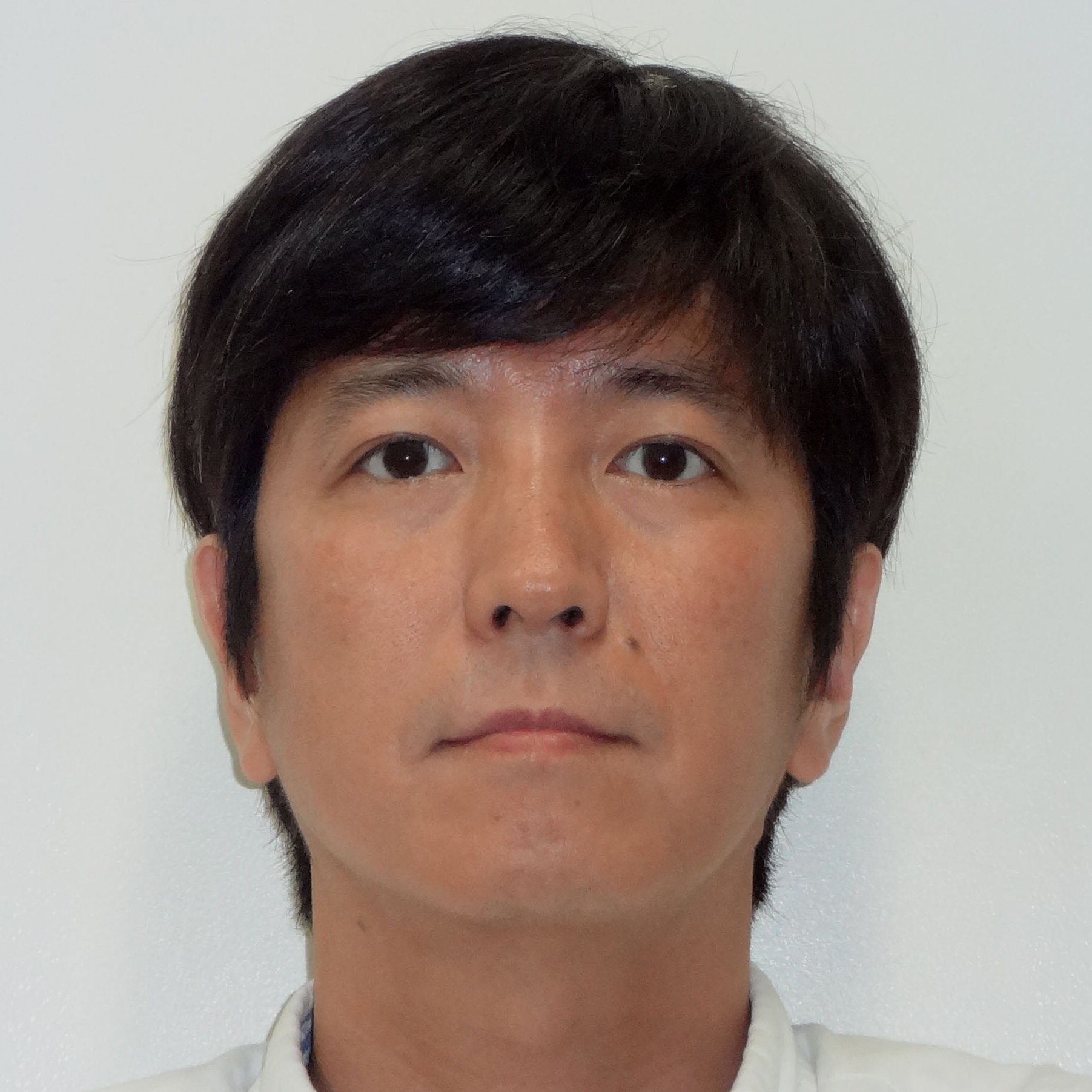
Prof. NISHIJIMA, Kazutoshi
Establishment and characterization of experimental animal models
Knowledge about animal characteristics and optimization of the breeding environment are indispensable for conducting scientifically meaningful animal experiments. Center for Animal Resources and Collaborative Study conducts the estblishment of experimental animal models and analysis of hteir characteristics, development of methods of strain preservation and breeding management suitable for each animal species (especially rabbits), from the viewpoint of veterinary and experimental animal sciences.
(※Currently, we do not accept students for trial enrollment.)
Article Information/Ggeneral remarks
Nishijima K, Kitajima S, Matsuhisa F, Niimi M, Wang C-c, Fan J (2021) Strategies for Highly Efficient Rabbit Sperm Cryopreservation. Animals. 2021 11: 1220.
Nishida Y, Nishijima K, Yamada Y, Tanaka H, Matsumoto A, Fan J, Uda Y, Tomatsu H, Yamamoto H, Kami K, Kitajima S, Tanaka K (2021) Whole-body insulin resistance and energy expenditure indices, serum lipids, and skeletal muscle metabolome in the lipoprotein lipase overexpression. Methabolomics 17: 26.
Matsuhisa F, Kitajima S, Nishijima K, Akiyoshi T, Masatoshi Morimoto M, Fan J (2020) Transgenic Rabbit Models: Now and the Future. Appl Sci 10: 7416.
Contact: NISHIJIMA, Kazutoshi TEL:+81-564-55-7781 / E-mail: kanish(at)nips.ac.jp
Lab Website
SUPPORTIVE CENTER FOR BRAIN RESEARCH, Section of Multiphoton Neuroimaging
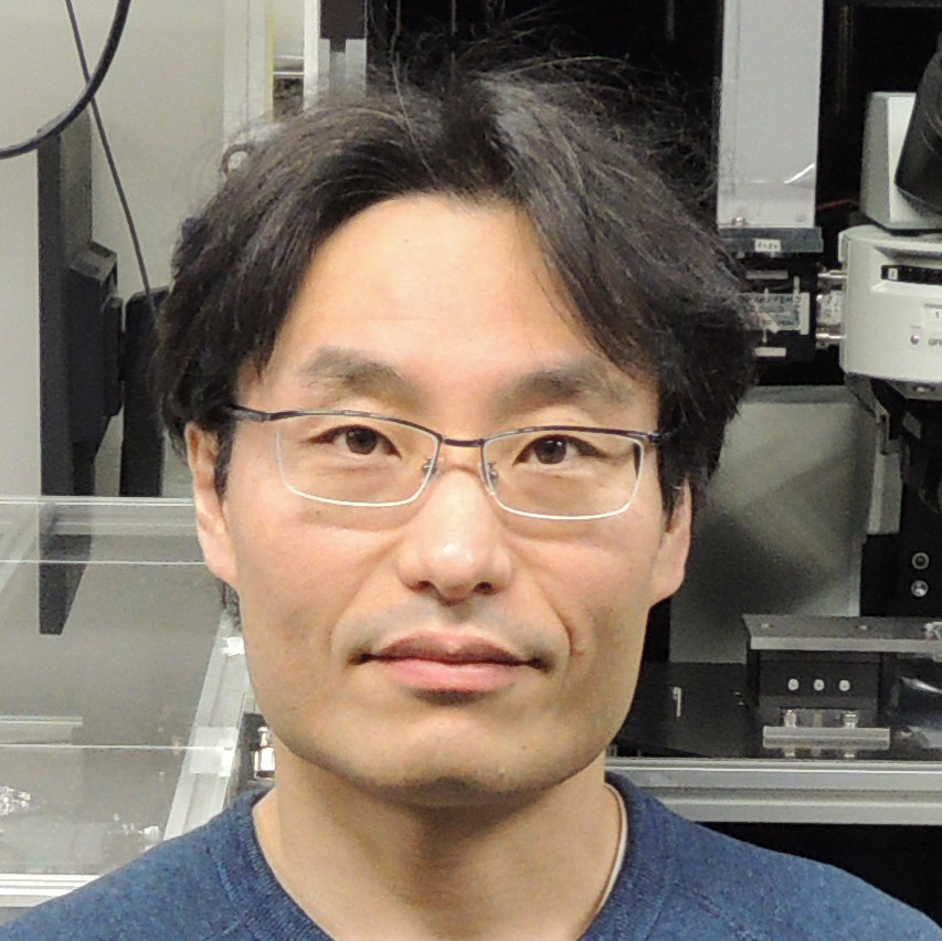
Assoc. Prof. MURAKOSHI, Hideji
Imaging signal transduction in subcellular structures of living cells by 2-photon fluorescence microscopy
We are working on imaging signal transduction in subcellular structure such as dendritic spine of hippocampal neurons using 2-photon fluorescence microscopy and fluorescence resonance energy transfer (FRET) techniques. By combining these techniques with optical manipulation techniques such as caged-compound uncaging and optogenetic tools, we are trying to understand the mechanism of physiological system such as memory system of brain at the level of single synapse.
Article Information/Ggeneral remarks
Ueda HH, Nagasawa Y, Sato A, Onda M, and Murakoshi H.* (2022) Chronic neuronal excitation leads to dual metaplasticity in the signaling for structural long-term potentiation. Cell Reports, 38(1), 110153.
Shibata AC, Ueda HH, Eto K, Onda M, Sato A, Ohba T, Nabekura J, and Murakoshi H* (2021) Photoactivatable CaMKII induces synaptic plasticity in single synapses. Nature communications 12, 751
Murakoshi H, Shin M, Parra-Bueno P, Szatmari EM, Shibata AC, and Yasuda R (2017) Kinetics of endogenous CaMKII required for synaptic plasticity revealed by optogenetic kinase inhibitor. Neuron 94: 37-47.
Murakoshi H, Wang H, Yasuda R (2011) Local, persistent activation of Rho GTPases during plasticity of single dendritic spines. Nature 472:100-104.
Contact: MURAKOSHI, Hideji Tel: +81-564-55-7857 / E-mail:murakosh(at)nips.ac.jp
Lab Website
SUPPORTIVE CENTER FOR BRAIN RESEARCH, Section of Brain Function Information
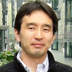
Prof. FUKUNAGA, Masaki
Imaging structure and function relationship of human brain by ultra- high field MRI
Magnetic resonance (MR) is an excellent technique for noninvasively observing living organisms' structure, function, metabolism, and molecular dynamics. In this section, we investigate the structure-function relationship of the human brain using 3 and 7-tesla ultra-high-field MR imaging and spectroscopy, and develop new imaging techniques to measure biological parameters. In addition, to understand the diseases through advanced brain imaging, we are participating in multi-center clinical research collaboration projects and promoting the exploration of endophenotypes and biomarkers of psychiatric disorders based on the analysis of big MR imaging data.
Article Information
Schijven D, Postema MC, Fukunaga M et al. (2023) Large-scale analysis of structural brain asymmetries in schizophrenia via the ENIGMA consortium. Proc Natl Acad Sci U S A. 120:e2213880120
Goda N, Hasegawa T, Koketsu D, Chiken S, Kikuta S, Sano H, Kobayashi K, Nambu A, Sadato N, Fukunaga M (2022) Cerebro-cerebellar interactions in nonhuman primates examined by optogenetic functional magnetic resonance imaging. Cereb Cortex Commun 3:tgac022
Maruyama S, Fukunaga M, Sugawara SK et al. (2021) Cognitive control affects motor learning through local variations in GABA within the primary motor cortex. Sci Rep 11:18566
Yamamoto T, Fukunaga M, Sugawara SK et al. (2021) Quantitative evaluations of geometrical distortion corrections in cortical surface‐based analysis of high‐resolution functional mri data at 7T. J Magn Reson Imaging. 53:1220
Contact: FUKUNAGA, Masaki TEL:+81-564-55-7842 / E-mail:fuku(at)nips.ac.jp
Lab Website
CENTER FOR GENETIC ANALYSIS OF BEHAVIOR, Section of Viral Vector Development
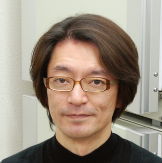
Assoc. Prof. KOBAYASHI, Kenta
Analysis of brain function using newly developed gene transfer system
To understand the mechanisms underlying higher brain functions, we need to analyze the roles of specific neuronal pathways forming the complex neural networks. We succeeded in developing a new gene transfer system to induce transgene expression in the specific neural pathway by using an adeno-associated viral vector and a novel type of lentiviral vector. We analyze the physiological functions of specific neural pathways forming cortico-basal ganglia circuits by using our newly developed gene transfer approach.
Article Information/Ggeneral remarks
Sano H, Kobayashi K, Yoshioka N, Takebayashi H, Nambu A (2020) Retrograde gene transfer into neural pathways mediated by adeno-associated virus (AAV)-AAV receptor interaction. J Neurosci Methods 345:108887.
Kobayashi K, Kato S, Kobayashi K (2018) Genetic manipulation of specific neural circuits by use of a viral vector system. J Neural Transm (Vienna) 125:67-75.
Kobayashi K, Inoue KI, Tanabe S, Kato S, Takada M, Kobayashi K (2017) Pseudotyped lentiviral vectors for retrograde gene delivery into target brain regions. Front Neuroanat 11:65.
Kobayashi K, Sano H, Kato S, Kuroda K, Nakamuta S, Isa T, Nambu A, Kaibuchi K, Kobayashi K (2016) Survival of corticostriatal neurons by Rho/Rho-kinase signaling pathway. Neurosci Lett 630:45-52.
Contact: KOBAYASHI, Kenta TEL:+81-564-55-7827 / E-mail:kobaya(at)nips.ac.jp
Lab Website
CENTER FOR GENETIC ANALYSIS OF BEHAVIOR, Section of Sensory Physiology

Assoc.Prof. SOKABE, Takaaki
Molecular mechanisms of sensing
We are clarifying molecular mechanisms of sensing such as thermosensation, nociception and taste sensation by focusing on TRP channels mainly with behavioral, electrophysiological, and molecular biological techniques. We are investigating fruit flies to seek novel mechanisms of temperature and mechanical sensations and developing new strategies of insect pest management by seeking novel repellents and insecticides.
Article Information
Ohnishi K, Sokabe T. (2024) Thermosensory Roles of G Protein-Coupled Receptors and Other Cellular Factors in Animals. Bioessays, e202400233.
Ohnishi K, Sokabe T, Miura T, Tominaga M, Ohta A, Kuhara A. (2024) G protein-coupled receptor-based thermosensation determines temperature acclimatization of Caenorhabditis elegans. Nat. Commun., 15:1660.
Sato S, Magaji AM, Tominaga M, Sokabe T. (2023) Avoidance of thiazoline compound depends on multiple sensory pathways mediated by TrpA1 and ORs in Drosophila. Front. Mol. Neurosci., 16:1249715.
Sokabe T, Bradshaw HB, Tominaga M, Leishman E, Chandel A, Montell C. (2022) Endocannabinoids produced in photoreceptor cells in response to light activate Drosophila TRP channels. Sci. Signal., 15(755):eabl6179.
Suito T, Nagao K, Juni N, Hara Y, Sokabe T, Atomi H, *Umeda M. (2022) Regulation of thermoregulatory behavior by commensal bacteria in Drosophila. Biosci. Biotechnol. Biochem., 86(8):1060-1070.
Contact: SOKABE, Takaaki TEL:+81-564-59-5287 / E-mail : sokabe(at)nips.ac.jp
Lab Website
Also, you can participate in visiting research divisions.
For the details of research activities, please contact the following
or visit our website. 〔
http://www.nips.ac.jp〕
National Institute for Physiological Sciences
SUPPORTIVE CENTER FOR BRAIN RESEARCH, Section of Brain Function Information , FUKUNAGA, Masaki
TEL:+81-564-55-7842 / E-mail:fuku(at)nips.ac.jp
 Assoc. Prof. TATEYAMA, Michihiro
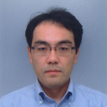 Prof. MURATA, Kazuyoshi
 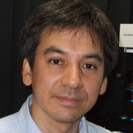 Prof. FURUSE, Mikio*
 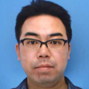 Assoc. Prof. IZUMI, Yasushi
 Prof. NISHIDA, Motohiro
  Assoc.Prof. NISHIMURA, Akiyuki
 Prof. MURAKAMI, Masaaki*
  Assoc.Prof. HASEBE, Rie
 Prof. KOBAYASHI, Toshihiro
  Prof. YOSHIMURA, Yumiko
  Prof. NEMOTO, Tomomi
  Assoc. Prof. ENOKI, Ryosuke
 Prof. WAKE, Hiroaki
  Prof. ISODA, Masaki
  Assoc.Prof. TOMATSU, Saeka
 Prof. KITAJO, Keiichi
  Prof. TAKEMURA, Hiromasa
  Prof. NISHIJIMA, Kazutoshi
 Assoc. Prof. MURAKOSHI, Hideji
  Prof. FUKUNAGA, Masaki
 Assoc. Prof. KOBAYASHI, Kenta
  Assoc.Prof. SOKABE, Takaaki